JAK1 (phospho Tyr1022) Polyclonal Antibody
- Catalog No.:YP0154
- Applications:IF;WB;IP;IHC;ELISA
- Reactivity:Human;Mouse;Rat
- Target:
- JAK1
- Fields:
- >>EGFR tyrosine kinase inhibitor resistance;>>PI3K-Akt signaling pathway;>>Necroptosis;>>Osteoclast differentiation;>>Signaling pathways regulating pluripotency of stem cells;>>NOD-like receptor signaling pathway;>>JAK-STAT signaling pathway;>>Th1 and Th2 cell differentiation;>>Th17 cell differentiation;>>Leishmaniasis;>>Toxoplasmosis;>>Tuberculosis;>>Hepatitis C;>>Hepatitis B;>>Measles;>>Human cytomegalovirus infection;>>Influenza A;>>Human papillomavirus infection;>>Human T-cell leukemia virus 1 infection;>>Kaposi sarcoma-associated herpesvirus infection;>>Herpes simplex virus 1 infection;>>Epstein-Barr virus infection;>>Coronavirus disease - COVID-19;>>Pathways in cancer;>>Viral carcinogenesis;>>Pancreatic cancer;>>PD-L1 expression and PD-1 checkpoint pathway in cancer
- Gene Name:
- JAK1
- Protein Name:
- Tyrosine-protein kinase JAK1
- Human Gene Id:
- 3716
- Human Swiss Prot No:
- P23458
- Mouse Swiss Prot No:
- P52332
- Immunogen:
- The antiserum was produced against synthesized peptide derived from human JAK1 around the phosphorylation site of Tyr1022. AA range:988-1037
- Specificity:
- Phospho-JAK1 (Y1022) Polyclonal Antibody detects endogenous levels of JAK1 protein only when phosphorylated at Y1022.
- Formulation:
- Liquid in PBS containing 50% glycerol, 0.5% BSA and 0.02% sodium azide.
- Source:
- Polyclonal, Rabbit,IgG
- Dilution:
- IF 1:50-200 WB 1:200 - 1:1000. IHC 1:100 - 1:300. ELISA: 1:10000. Not yet tested in other applications.
- Purification:
- The antibody was affinity-purified from rabbit antiserum by affinity-chromatography using epitope-specific immunogen.
- Concentration:
- 1 mg/ml
- Storage Stability:
- -15°C to -25°C/1 year(Do not lower than -25°C)
- Other Name:
- JAK1;JAK1A;JAK1B;Tyrosine-protein kinase JAK1;Janus kinase 1;JAK-1
- Observed Band(KD):
- 132kD
- Background:
- This gene encodes a membrane protein that is a member of a class of protein-tyrosine kinases (PTK) characterized by the presence of a second phosphotransferase-related domain immediately N-terminal to the PTK domain. The encoded kinase phosphorylates STAT proteins (signal transducers and activators of transcription) and plays a key role in interferon-alpha/beta and interferon-gamma signal transduction. Alternative splicing results in multiple transcript variants. [provided by RefSeq, Mar 2016],
- Function:
- catalytic activity:ATP + a [protein]-L-tyrosine = ADP + a [protein]-L-tyrosine phosphate.,domain:Possesses two phosphotransferase domains. The second one probably contains the catalytic domain (By similarity), while the presence of slight differences suggest a different role for domain 1.,domain:The FERM domain mediates interaction with JAKMIP1.,function:Tyrosine kinase of the non-receptor type, involved in the IFN-alpha/beta/gamma signal pathway. Kinase partner for the interleukin (IL)-2 receptor.,sequence caution:Translation N-terminally extended.,similarity:Belongs to the protein kinase superfamily. Tyr protein kinase family. JAK subfamily.,similarity:Contains 1 FERM domain.,similarity:Contains 1 protein kinase domain.,similarity:Contains 1 SH2 domain.,subcellular location:Wholly intracellular, possibly membrane associated.,subunit:Interacts with IL31RA, JAKMIP1 and SHB.,tissue specif
- Subcellular Location:
- Endomembrane system; Peripheral membrane protein. Wholly intracellular, possibly membrane associated.
- Expression:
- Expressed at higher levels in primary colon tumors than in normal colon tissue. The expression level in metastatic colon tumors is comparable to the expression level in normal colon tissue.
Glycogen metabolism regulates macrophage-mediated acute inflammatory responses. Nature Communications Nat Commun. 2020 Apr;11(1):1-16 WB Mouse 1:1000 BMDMs
Treadmill Exercise Attenuates Cerebral Ischemia–Reperfusion Injury by Promoting Activation of M2 Microglia via Upregulation of Interleukin-4. Frontiers in Cardiovascular Medicine Front Cardiovasc Med. 2021 Oct;0:1282 WB Rat 1:500 Penumbra
Herpes Simplex Virus 1 UL36USP Antagonizes Type I Interferon-Mediated Antiviral Innate Immunity. JOURNAL OF VIROLOGY J Virol. 2018 Oct;: IP Human HEK293T cell
Paeoniflorin‐6′‐o‐benzene sulfonate (CP‐25) improves vasculitis through inhibiting IL‐17A/JAK/STAT3 signaling pathway in endothelial cells of HFD CIA rats. PHYTOTHERAPY RESEARCH Phytother Res. 2021 Feb;35(2):1033-1047 WB Human 1 : 500 HUVECs
C6orf120 gene knockout in rats mitigates concanavalin A‑induced autoimmune hepatitis via regulating NKT cells. CELLULAR IMMUNOLOGY Cell Immunol. 2022 Jan;371:104467 WB Rat Liver
Novel paeonol derivatives: Design, synthesis and anti-inflammatory activity in vitro and in vivo. BIOORGANIC CHEMISTRY Bioorg Chem. 2020 May;98:103735 WB Mouse RAW264.7 cell
朱悦, et al. "芍药苷-6′-O-苯磺酸酯通过调节 JAK/STAT3RORγ-t/IL-17A 信号通路抑制人外周血 Th17 细胞的分化." (2020).
Alpha-Momorcharin Inhibits Proinflammatory Cytokine Expression by M1 Macrophages but Not Anti-Inflammatory Cytokine Expression by M2 Macrophages Journal of Inflammation Research Fubing Shen WB Human
Phillygenin inhibited M1 macrophage polarization and reduced hepatic stellate cell activation by inhibiting macrophage exosomal miR-125b-5p BIOMEDICINE & PHARMACOTHERAPY Cheng Ma WB Mouse RAW264.7 cells
- June 19-2018
- WESTERN IMMUNOBLOTTING PROTOCOL
- June 19-2018
- IMMUNOHISTOCHEMISTRY-PARAFFIN PROTOCOL
- June 19-2018
- IMMUNOFLUORESCENCE PROTOCOL
- September 08-2020
- FLOW-CYTOMEYRT-PROTOCOL
- May 20-2022
- Cell-Based ELISA│解您多样本WB检测之困扰
- July 13-2018
- CELL-BASED-ELISA-PROTOCOL-FOR-ACETYL-PROTEIN
- July 13-2018
- CELL-BASED-ELISA-PROTOCOL-FOR-PHOSPHO-PROTEIN
- July 13-2018
- Antibody-FAQs
- Products Images
.jpg)
- Ma, J., Wei, K., Liu, J. et al. Glycogen metabolism regulates macrophage-mediated acute inflammatory responses. Nat Commun 11, 1769 (2020).
-if-60.jpg)
- Immunofluorescence analysis of mouse-liver tissue. 1,JAK1 (phospho Tyr1022) Polyclonal Antibody(red) was diluted at 1:200(4°C,overnight). 2, Cy3 labled Secondary antibody was diluted at 1:300(room temperature, 50min).3, Picture B: DAPI(blue) 10min. Picture A:Target. Picture B: DAPI. Picture C: merge of A+B
-if-61.jpg)
- Immunofluorescence analysis of mouse-liver tissue. 1,JAK1 (phospho Tyr1022) Polyclonal Antibody(red) was diluted at 1:200(4°C,overnight). 2, Cy3 labled Secondary antibody was diluted at 1:300(room temperature, 50min).3, Picture B: DAPI(blue) 10min. Picture A:Target. Picture B: DAPI. Picture C: merge of A+B
poly-ihc-human-liver-cancer.jpg)
- Immunohistochemical analysis of paraffin-embedded Human-liver-cancer tissue. 1,JAK1 (phospho Tyr1022) Polyclonal Antibody was diluted at 1:200(4°C,overnight). 2, Sodium citrate pH 6.0 was used for antibody retrieval(>98°C,20min). 3,Secondary antibody was diluted at 1:200(room tempeRature, 30min). Negative control was used by secondary antibody only.
poly-ihc-human-stomach-cancer.jpg)
- Immunohistochemical analysis of paraffin-embedded Human-stomach-cancer tissue. 1,JAK1 (phospho Tyr1022) Polyclonal Antibody was diluted at 1:200(4°C,overnight). 2, Sodium citrate pH 6.0 was used for antibody retrieval(>98°C,20min). 3,Secondary antibody was diluted at 1:200(room tempeRature, 30min). Negative control was used by secondary antibody only.
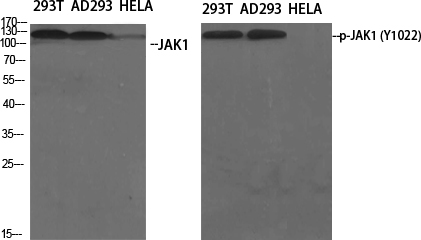
- Western Blot analysis of various cells using Phospho-JAK1 (Y1022) Polyclonal Antibody diluted at 1:500
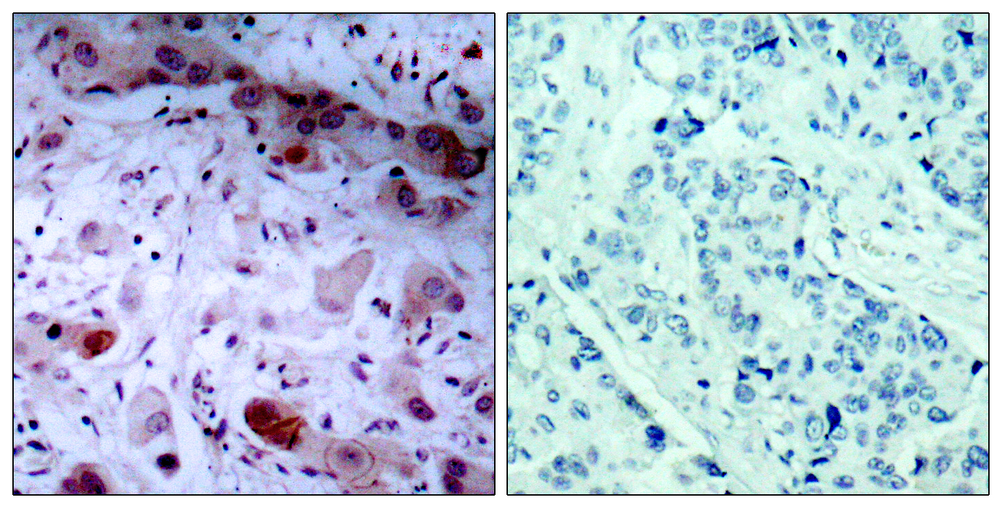
- Immunohistochemistry analysis of paraffin-embedded human breast carcinoma, using JAK1 (Phospho-Tyr1022) Antibody. The picture on the right is blocked with the phospho peptide.
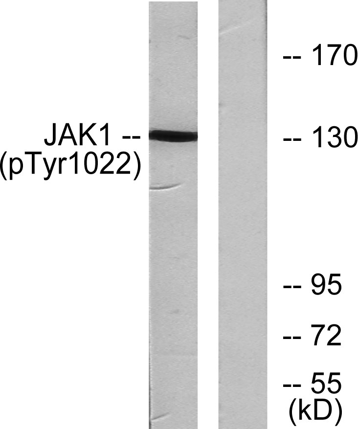
- Western blot analysis of lysates from A549 cells , using JAK1 (Phospho-Tyr1022) Antibody. The lane on the right is blocked with the phospho peptide.


