HP-1α mouse Monoclonal Antibody(5E3)
- Catalog No.:YM3744
- Applications:IF;WB;IHC
- Reactivity:Human;Mouse;Rat
- Target:
- HP1α
- Gene Name:
- CBX5 HP1A
- Protein Name:
- Chromobox protein homolog 5 (Antigen p25) (Heterochromatin protein 1 homolog alpha) (HP1 alpha)
- Human Gene Id:
- 23468
- Human Swiss Prot No:
- P45973
- Mouse Swiss Prot No:
- Q61686
- Immunogen:
- Recombinant Protein of HP-1α
- Specificity:
- The antibody detects endogenous HP-1α protein
- Formulation:
- Liquid in PBS containing 50% glycerol, 0.5% BSA and 0.02% sodium azide.
- Source:
- Monoclonal, Mouse
- Dilution:
- IF 1:50-200 WB 1:500-2000,IHC 1:50-300
- Purification:
- The antibody was affinity-purified from mouse antiserum by affinity-chromatography using epitope-specific immunogen.
- Concentration:
- 1 mg/ml
- Storage Stability:
- -15°C to -25°C/1 year(Do not lower than -25°C)
- Other Name:
- Chromobox protein homolog 5 (Antigen p25) (Heterochromatin protein 1 homolog alpha) (HP1 alpha)
- Observed Band(KD):
- 22kD
- Background:
- This gene encodes a highly conserved nonhistone protein, which is a member of the heterochromatin protein family. The protein is enriched in the heterochromatin and associated with centromeres. The protein has a single N-terminal chromodomain which can bind to histone proteins via methylated lysine residues, and a C-terminal chromo shadow-domain (CSD) which is responsible for the homodimerization and interaction with a number of chromatin-associated nonhistone proteins. The encoded product is involved in the formation of functional kinetochore through interaction with essential kinetochore proteins. The gene has a pseudogene located on chromosome 3. Multiple alternatively spliced variants, encoding the same protein, have been identified. [provided by RefSeq, Jul 2008],
- Function:
- function:Component of heterochromatin. Recognizes and binds histone H3 tails methylated at 'Lys-9', leading to epigenetic repression. Can interact with lamin B receptor (LBR). This interaction can contribute to the association of the heterochromatin with the inner nuclear membrane. Involved in the formation of functional kinetochore through interaction with MIS12 complex proteins.,PTM:Phosphorylation of HP1 and LBR may be responsible for some of the alterations in chromatin organization and nuclear structure which occur at various times during the cell cycle (By similarity). Phosphorylated during interphase and possibly hyper-phosphorylated during mitosis.,similarity:Contains 2 chromo domains.,subcellular location:Component of centromeric and pericentromeric heterochromatin. Associates with chromosomes during mitosis. Associates specifically with chromatin during metaphase and anaphase.,
- Subcellular Location:
- Nucleus . Chromosome . Chromosome, centromere . Colocalizes with HNRNPU in the nucleus (PubMed:19617346). Component of centromeric and pericentromeric heterochromatin. Associates with chromosomes during mitosis. Associates specifically with chromatin during metaphase and anaphase. .
- Expression:
- Epithelium,Fetal brain cortex,Placenta,
- June 19-2018
- WESTERN IMMUNOBLOTTING PROTOCOL
- June 19-2018
- IMMUNOHISTOCHEMISTRY-PARAFFIN PROTOCOL
- June 19-2018
- IMMUNOFLUORESCENCE PROTOCOL
- September 08-2020
- FLOW-CYTOMEYRT-PROTOCOL
- May 20-2022
- Cell-Based ELISA│解您多样本WB检测之困扰
- July 13-2018
- CELL-BASED-ELISA-PROTOCOL-FOR-ACETYL-PROTEIN
- July 13-2018
- CELL-BASED-ELISA-PROTOCOL-FOR-PHOSPHO-PROTEIN
- July 13-2018
- Antibody-FAQs
- Products Images
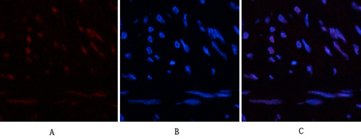
- Immunofluorescence analysis of human-uterus tissue. 1,HP-1α Mouse Monoclonal Antibody(5E3)(red) was diluted at 1:200(4°C,overnight). 2, Cy3 labled Secondary antibody was diluted at 1:300(room temperature, 50min).3, Picture B: DAPI(blue) 10min. Picture A:Target. Picture B: DAPI. Picture C: merge of A+B
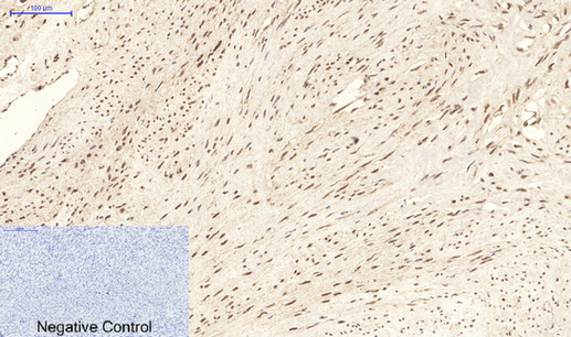
- Immunohistochemical analysis of paraffin-embedded Human-uterus tissue. 1,HP-1α Mouse Monoclonal Antibody(5E3) was diluted at 1:200(4°C,overnight). 2, Sodium citrate pH 6.0 was used for antibody retrieval(>98°C,20min). 3,Secondary antibody was diluted at 1:200(room tempeRature, 30min). Negative control was used by secondary antibody only.
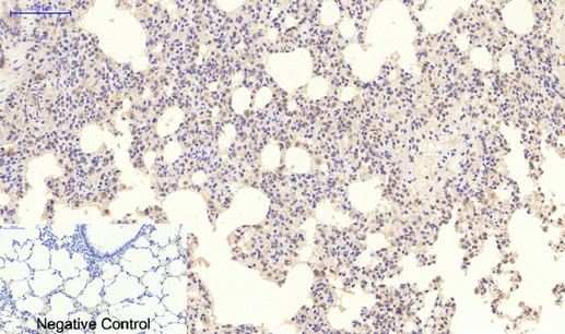
- Immunohistochemical analysis of paraffin-embedded Rat-lung tissue. 1,HP-1α Mouse Monoclonal Antibody(5E3) was diluted at 1:200(4°C,overnight). 2, Sodium citrate pH 6.0 was used for antibody retrieval(>98°C,20min). 3,Secondary antibody was diluted at 1:200(room tempeRature, 30min). Negative control was used by secondary antibody only.
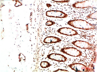
- Immunohistochemical analysis of paraffin-embedded Human Colon Carcinoma Tissue using HP-1 α Mouse mAb diluted at 1:200
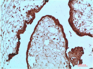
- Immunohistochemical analysis of paraffin-embedded Human Placenta Tissue using HP-1 α Mouse mAb diluted at 1:200
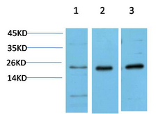
- Western blot analysis of 1) Hela Cell Lysate, 2)3T3 Cell Lysate, 3) PC12 Cell Lysate using HP-1γα Mouse mAb diluted at 1:1000.


